Try Now
One platform to teach, diagnose, and discover— without expensive scanners, desktop workstations, or steep learning curves.
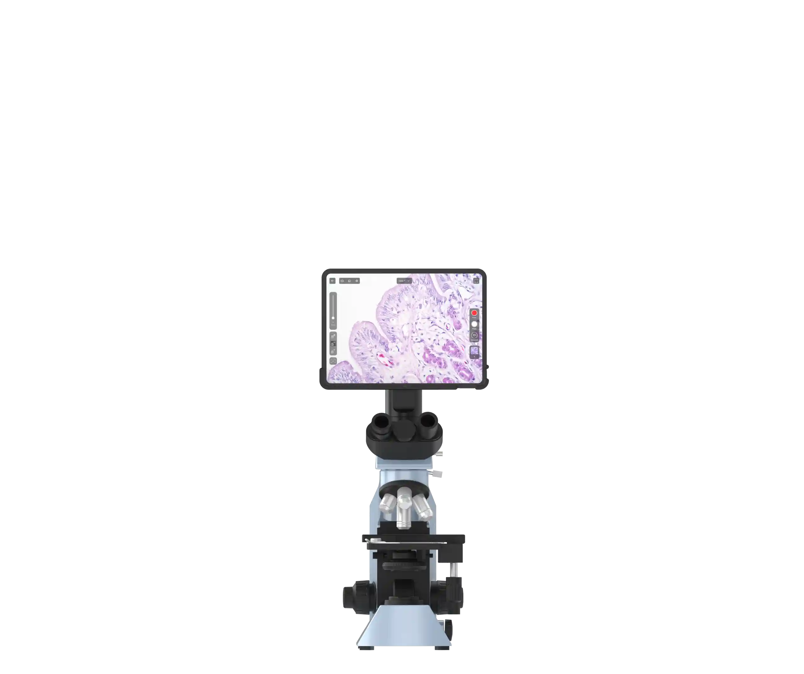
The Microscope has evolved.
Meet its next stage: intelligent, connected, effortless.


Voice-to-record in real time — no keyboard, no lag.
Dictate in your own words; Eva captures your observations the moment you make them. It intelligently handles complex medical terms and places your notes directly into the case record.
Build Once. Report Perfectly, Every Time.
Create your ideal report templates with custom branding, synoptic tables, and required fields. Share and lock them in a single click, instantly equipping your entire team with a tool for consistent, high-quality reporting. It’s the simplest way to standardize excellence.

The Consult is in the Case.
Need a second opinion? Call a remote pathologist or clinician directly within Eva. Our platform doesn't just connect you; it listens. Eva automatically transcribes the entire conversation and appends it to the case file, creating a complete, auditable record of the consult. No more lost notes—just clear, collaborative decisions.
Manage Organisation & Control Access with Roles
Assign granular permissions to pathologists, technicians, and admin staff to ensure data security.

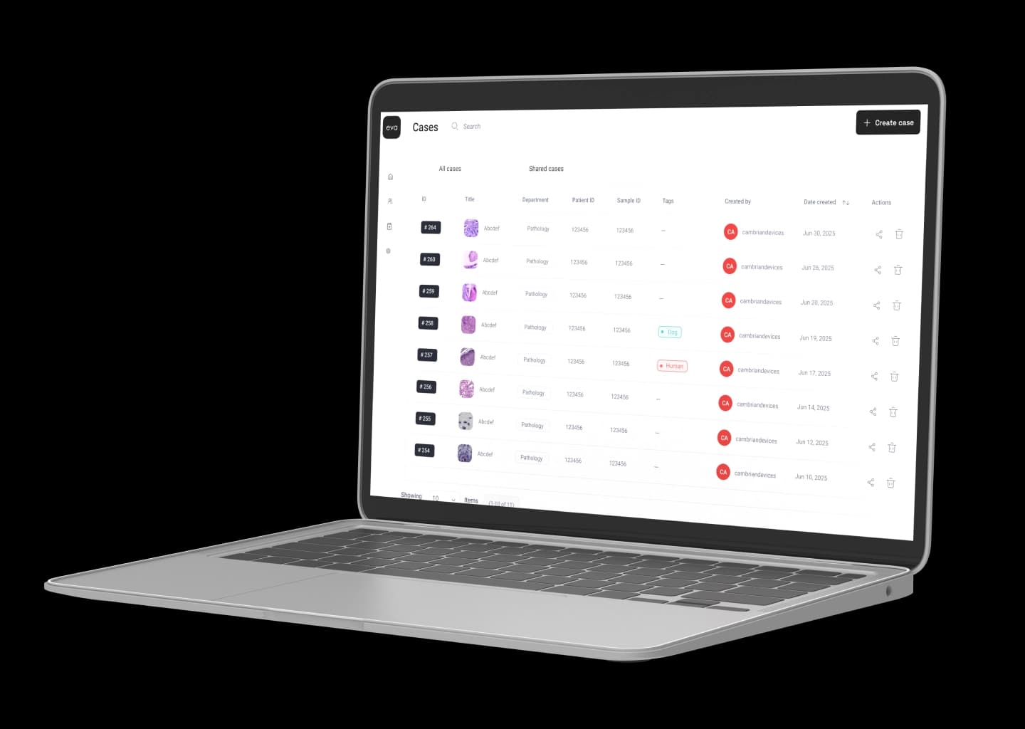
Organise Cases
Store cases with metadata and all media to keep high volumes tidy and findable for clean, efficient workflow.

Tag with Precision (Multi-Level Tagging)
Apply detailed tags at specimen, stain, and lesion levels for lightning-fast search and retrieval.
Integrate with LIMS & HIS
Seamlessly pull patient demographics and order info, and push finalized reports back into your lab or hospital system.
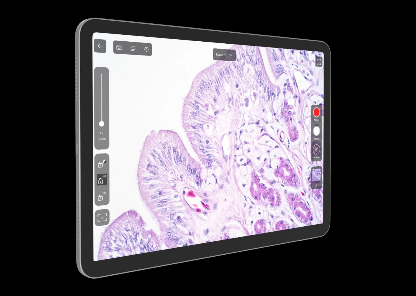
Capture High-Res Images
Save publication-quality images directly from your field of view, preserving every critical detail at sub-micron resolution.

Enhance Diagnostic Visuals
Instantly modify brightness, contrast, and color balance to clarify subtle morphological features.
Annotate & Label Findings
Draw shapes, place arrows, and add structured labels to any region of interest for clear documentation and communication.

Count Cells with Grids
Overlay a configurable grid for rapid, consistent, and reproducible cell counting.

Measure with Precision
Use automatically calibrated tools to measure length, area, and cellular structures with sub-micron accuracy.

Dictate Your Notes
Use medical grade voice-to-text to capture observations hands-free while you navigate the slide, speeding up your workflow.
Discuss Findings In-Context
Pin comments directly onto specific image regions to facilitate clear, real-time discussions with colleagues.

Share Cases for Consultation
Securely share cases with specialists, colleagues, or students for rapid feedback and second opinions.

Launch Tele-Consults
Initiate a secure telepathology session (via Google Meet) directly from the case view. Consult live and receive an automatic transcription for your records.
Export with Ease
One-click export of images and video clips to PNG, JPEG, or MP4. Share directly via email, WhatsApp, or secure link.

Build Custom Reports
Design and generate patient and synoptic reports (surgical, cytology, IHC) that match your organization's precise formatting needs.

Ensure Full Traceability
Maintain a complete history of all user activity on a case, perfect for regulatory or accreditation audits.


Canine skin — melanoma (H&E), brightfield, 10×

Cervical cytology — squamous cell carcinoma (Pap), brightfield, 63×

Canine blood smear — microfilariae (Giemsa), brightfield, 100×

Breast carcinoma — cytokeratin 5/6 (IHC), brightfield, 10×

Sputum smear — Mycobacterium (auramine–rhodamine), fluorescence

Douglas fir fibers — phase-contrast micrograph (stained)

Necrotizing fasciitis tissue — Gram-positive diplococci (Gram stain), brightfield, 40×
Leptospira — live wet mount, dark-field microscopy

Urine sediment — granular ('muddy brown') casts (acute tubular necrosis), brightfield,
Available now
on web
and android
Will Eva fit my microscope?
+Eva mounts to the trinocular head on Olympus CX23/33/43, BX43/53, and Magnus MX series microscopes. If yours isn’t supported, we offer an Eva + Magnus MX35i Pro bundle; our compatibility list is growing—check back or contact us to confirm your model.
Can I still use the eyepieces?
+Yes, Eva uses the trinocular port, so you can look through the binoculars while the screen shows the same view (par-focal) for easy back-and-forth.
Will the digital image match what I see by eye?
+Yes—the tablet view is par-focal with your eyepieces (same magnification and FOV), and the live feed tracks stage motion with zero lag.
Can I create whole-slide images?
+Today you can capture high-resolution photos and 4K videos of your field of view; manual stitching for virtual slides is on the roadmap.
How do I share images or cases?
+Invite collaborators by secure email link (they sign in to view under “Shared with me”); export PNG/JPEG/MP4 or share a secure link/WhatsApp—subject to your organisation’s policy.